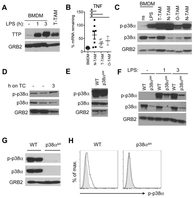
Figure 5. Reduced TTP-dependent mRNA decay coincides with chronic p38α activation in TAMs and PAMs.
A, TTP expression analyzed by immunoblotting of whole cell lysates of EG7 TAMs and resting or LPS treated BMDMs. Blots were re-probed using anti-GRB2 antibody to confirm equal loading and represent 1 out of 3 experiments. B, TNF mRNA stability determined by qRT-PCR by comparing cells before and 90 minutes after transcriptional blockade by Actinomycin D in BMDMs and TAMs isolated from EG7 thymomas (T-TAM), neuroblastomas (N-TAM) or osteosarcomas (O-TAM). Values were normalized to GAPDH and represent % TNF mRNA remaining after Actinomycin D treatment as mean and SEM of at least 2 experiments (n ≥ 2). C, immunoblot analysis of total cell lysates of non-stimulated (ns) or 1hr LPS stimulated (LPS) BMDMs as well as TAMs isolated from different tumor models as in B, probed with anti-phospho-p38, anti-p38 or anti-GRB2 as loading control. The blot represents 1 out of 3 experiments. D, p38 phosphorylation analyzed by immunoblotting of whole EG7 TAM lysates from WT TAMs directly after isolation (−) or after resting them on tissue culture dishes (TC) for 3 hrs. E and F, immunoblot analysis of whole EG7 TAM lysates (E) or BMDMs left untreated or stimulated with LPS for 1 and 3 hrs (F) isolated from WT or p38αΔM animals. Blots represent 1 out of 2 experiments. G, Whole EG7 TAM lysates from WT or p38αΔH animals as in (E) analyzed for p38 and p-p38 protein expression by immunoblotting (n = 2). H, flow cytometry analysis of p-p38 in CD11b+ EG7 TAMs isolated from WT or p38αΔH animals (n ≥ 4). Representative plots are shown with unstained control as grey line.

