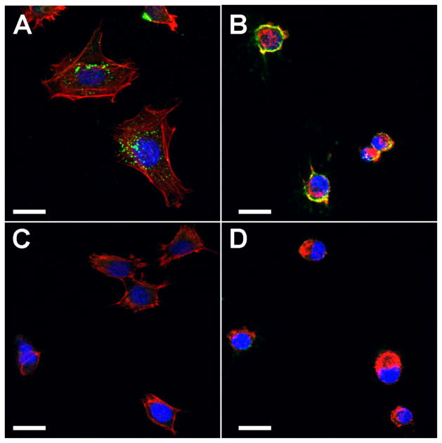
Figure 5. Cytochalasin D treatment prevents internalization of antibodies by SCA9-15 cells.
Confocal images show that cells incubated with Ro52-immune sera have cytoplasmic IgG (green) staining (A), which is not seen in cells incubated with control sera (C). Cytochalasin D treatment localizes antibody reactivity to the surface (B). Cells treated with Cytochalasin D, become rounded due to collapse in actin filaments (red) stained with Phalloidin Alexa Fluor 568. Similar results were obtained in 3 additional experiments. Scale bar = 10μm.

