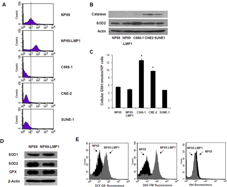
Fig 1. Oncogenic transformation by LMP1 causes increased ROS generation.
A: Basal hydrogen peroxide levels in immortalized nasopharyngeal epithelial cells (NP69 and NP69-LMP1) and NPC cells (C666-1, CNE-2 and SUNE-1) were detected by flow cytometry using DCF-DA. Each histogram is representative of three experiments. Compared to NP69 cells, NP69-LMP1 cells exhibit significantly higher level of hydrogen peroxide (p<0.05). B: Basal protein expression of catalase and superoxide dismutase 2 (SOD2) in immortalized nasopharyngeal epithelial cells (NP69 and NP69-LMP1) and NPC cells (C666-1, CNE-2 and SUNE-1). β-Actin served as a loading control. C: Comparison of total cellular GSH in immortalized nasopharyngeal epithelial cells (NP69 and NP69-LMP1) and NPC cells (C666-1, CNE-2 and SUNE-1) (mean ± SD of three experiments, * p<0.05). D: Basal protein expression of catalase, glutathione peroxidase (GPX) and superoxide dismutase (SOD1 and SOD2) in NP69 and NP69-LMP1 cells. β-Actin served as a loading control. E: Increase in ROS levels in NP69-LMP1 cells detected by flow cytometry using DCF-DA (left panel) and DAF-FM (middle panel). Superoxide was detected using HEt (right panel). Each histogram is representative of three experiments. Compared to NP69 cells, NP69-LMP1 cells exhibit significantly higher level of ROS contents, including hydrogen peroxide and nitrogen oxide (p<0.05).

