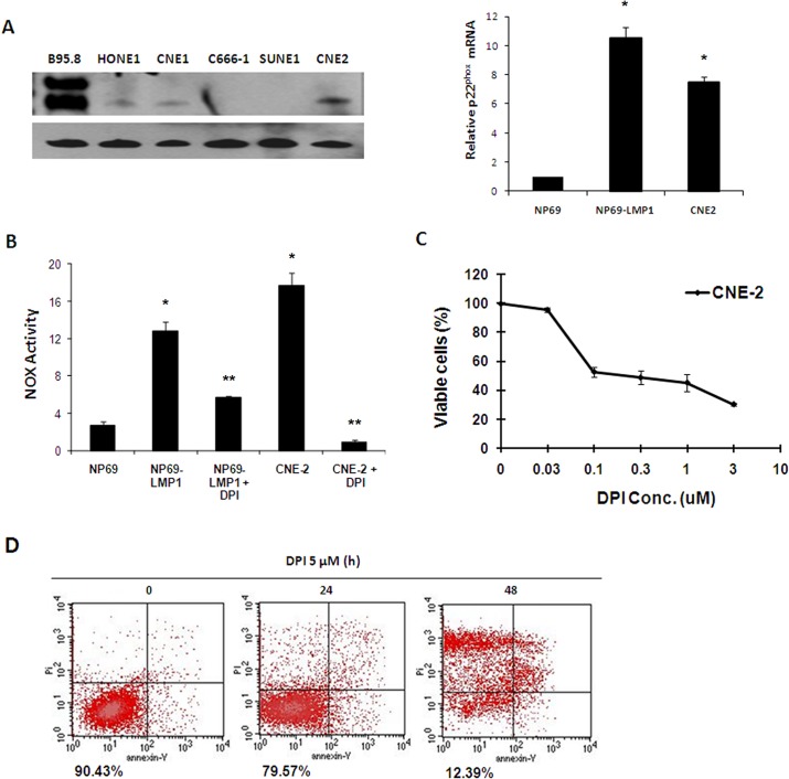
Fig 6. NOX activation makes NPC cells vulnerable to the NOX inhibitor DPI.
A: p22phox mRNA expression level was evaluated in NP69, NP69-LMP1 and CNE2 cells by RT-PCR and real-time quantitative PCR. LMP1 expression was detected in NPC cells and B95.8 B lymphoma cells. β-Actin served as a loading control (mean ± SD of three experiments). Compared to NP69 cells, NP69-LMP1 and CNE2 cells had significantly higher p22phox mRNA expression level (*p<0.01). B: Comparison of NOX activity and the effect of DPI on NOX activity in NP69, NP69-LMP1 and CNE2 cells, measured by a luminometer using lucigenin in the presence of NADPH (mean ± SD of three experiments). Compared to NP69 cells, NP69-LMP1 and CNE2 cells had significantly higher NOX activity (* p < 0.01). DPI treatment could significantly suppress NOX activity in NP69-LMP1 and CNE2 cells (** p<0.01). C: Mortality effect of 0.03–10 μM DPI on CNE2 NPC cells, evaluated using an MTT assay (mean ± SD of three experiments). DPI suppressed CNE2 cells proliferation. D: Mortality effect of 5 μM DPI on CNE2 cells, detected using annexin V-PI staining and flow cytometry. The numbers under the plots indicate the live cells with both low annexin V staining and low PI staining. Each histogram is representative of three experiments.

