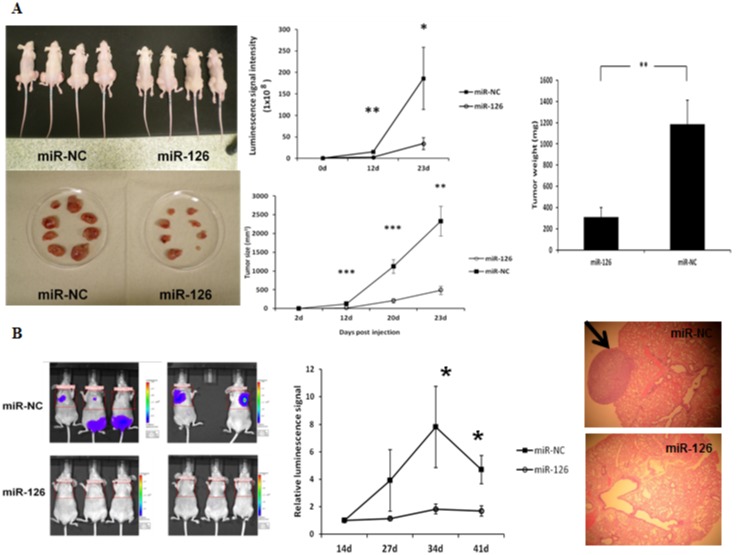
Fig 4. MiR-126-3p overexpression inhibits tumor growth and tumor metastasis in vivo.
(A) Growth of tumor xenografts in nude mice. Left panel: representative images of mice xenograft size at autopsy. Middle and right panel: tumor luciferase activity and tumor volume measurement, and weight. FTC-133-Luc2 cells transfected with miR-126-3p and miR-NC were inoculated subcutaneously in the flanks of athymic nude mice. (B) Tumor metastasis. Left panel: Representative images of mice with metastases showing luminescence signal. Middle panel: Quantification of luminescence signal intensity differences between miR-126-3p and miR-NC. FTC-133-Luc2 cells transfected with miR-126-3p and miR-NC were injected into athymic nude mice via the tail vein, and the mice were imaged with a Xenogen IVIS 100 system. The relative luminescence signal of each mouse is calculated as the ratio of original signal to the signal taken 14 days post-injection. The images shown here were taken 7 weeks after vein injection of tumor cells. Error bars represent SEM (* indicates p<0.05; ** indicates p<0.01; *** indicates p<0.001). All animal experiments were repeated twice. Right panel: A representative microscopic image (hematoxylin and eosin [H&E] staining) of metastatic lung tumor induced by FTC-133-Luc2 cells transfected with miR-NC and an H&E-stained section of metastatic lung tumor induced by FTC-133-Luc2 cells transfected with miR-126-3p.

