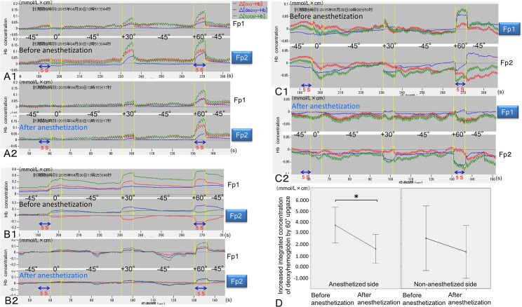
Fig 6. Impaired activation of the ipsilateral ventromedial prefrontal cortex by unilateral anesthetization of mechanoreceptors in the supratarsal Müller muscle.
Relative changes in the deoxyhemoglobin, oxyhemoglobin, total hemoglobin concentrations (mmol/L × cm) were measured at Fp1 and Fp2 according to the changes in the degree of eyelid retraction such as 45° downgaze (-45°), primary gaze (0°), 30° upgaze (+30°), and 60° upgaze (+60°) before (A1, B1, and C1) and after (A2, B2, and C2) anesthetization. Before anesthetization, three representative examples show bilateral activation (A1), asymmetrical bilateral activation (B1), and unilateral activation and contralateral deactivation for 5 s of 60° upgaze (+60°) (C1). After anesthetization, all of the examples show impaired activation at Fp1 or Fp2 on the anesthetized side (A2, B2, and C2). (D) On the anesthetized side, the increased integrated concentration of deoxyhemoglobin by 60° upgaze for 5 s after anesthetization was significantly smaller than that before anesthetization (*P < 0.01). However, on the non-anesthetized side, the increased integrated concentration of deoxyhemoglobin by 60° upgaze for 5 s after anesthetization was not significantly smaller than that before anesthetization.

