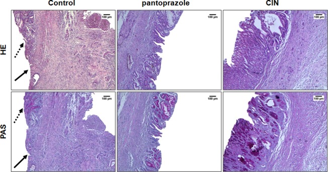
Fig 3. Photomicrographs of gastric mucosa stained with HE and PAS of the rats subjected to induction of chronic ulcer by 30% acetic acid.

Animals were treated orally with 1% Tween-80 aqueous solution (control group), pantoprazole (40 mg/kg) or CIN (100 mg/kg) for 14 days. The filled arrow indicates the absence of the epithelial layer (ulcer area internal) and the dashed arrow indicates epithelial layer remaining (ulcer edge). Haematoxylin/eosin (HE) and Periodic Acid–Schiff staining (PAS), magnification, 40x.
