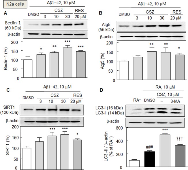
Fig 2. Increases in the expressions of beclin-1 (A), Atg5 (B), and SIRT1 protein (C) by cilostazol (CSZ, 3–30 μM; incubation for 3 h) and resveratrol (RES, 20 μM) in the presence of exogenous Aβ1–42 (10 μM) in N2a cells. D. Enhancement of LC3-II levels in the culture media containing 10 μM retinoic acid by cilostazol (10 μM), and its blockade by 3-methyladenine (3-MA, 2.5 mM).

Means ± SDs are expressed as percentages of DMSO (vehicle) or absence of retinoic acid (RA-) (N = 4). ### P < 0.001, RA−; *P < 0.05, **P < 0.01, ***P < 0.001 vs. DMSO; ††† P < 0.001 vs. 10 μM cilostazol.
