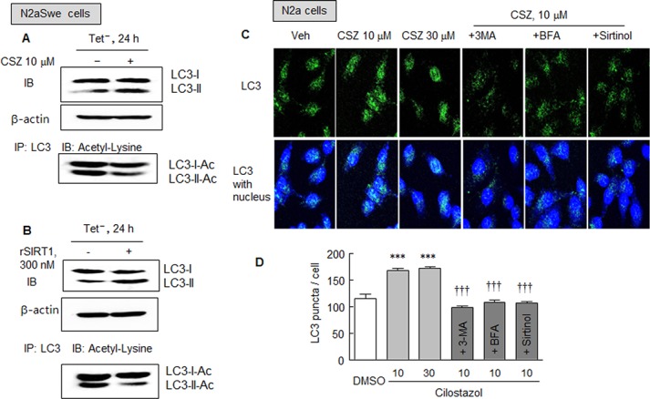
Fig 6. A and D. Immunoprecipitation analysis. Whole cell lysates were obtained from N2aSwe cell lysates that were cultured in Tet- condition for 24 h with or without cilostazol (10 μM, A) or rSIRT1 (recombinant SIRT1, 300 nM; B). Upper panels (A and B): the effects of cilostazol and rSIRT1 on LC3-II expression were confirmed. Lower panels (A and B): cell lysates were immunoprecipitated with LC-3 antibody and then immunoblotted for acetylated LC3 LC3-1/II-Ac) using an anti-acetyl lysine antibody. The blot shown is representative of three experiments that produced similar results. C. Immunofluorescent assay of LC3 puncta in N2a cells treated with or without cilostazol (10 and 30 μM) after being pretreated with 3-methyladenine (2.5 mM), bafilomycin A1 (100 nM) or sirtinol (20 μM). D. Quantitative analysis was performed by counting numbers of LC3 puncta/cell. Results are the means ± SDs of 4 experiments.

***P < 0.001 vs. DMSO. ††† P < 0.001 vs. cilostazol (10 μM).
