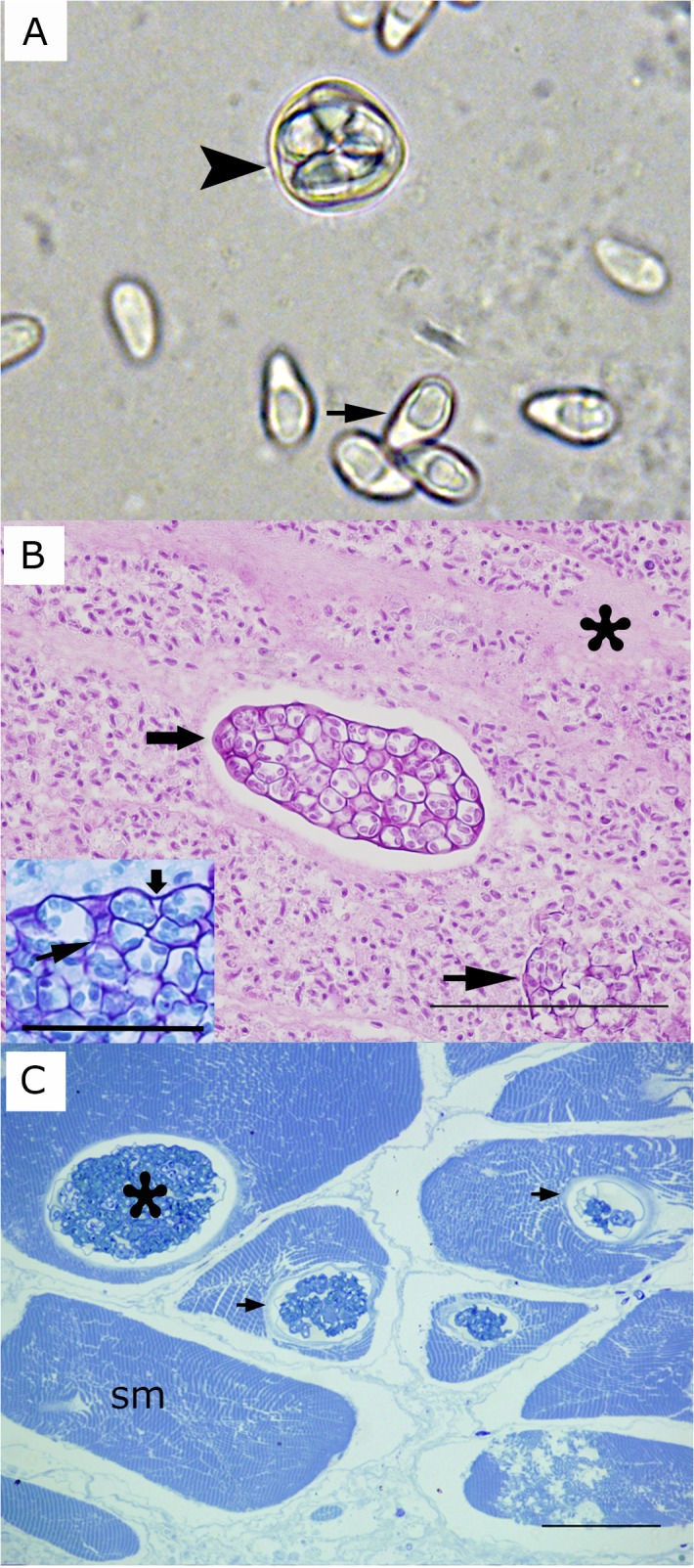
Fig 2.

A) Fresh preparation of microsporidia-infected fish. Arrow indicates a spore of Heterosporis sutherlandae n. sp. Arrowhead shows a sporophorous vesicle with several spores. B) Widespread muscle destruction due to H. sutherlandae. Mature spores have replaced muscle cells and are surrounded by loose fibrous tissue (asterisk). The wide arrow indicates a large sporophorocyst containing sporoblast and spores. The narrow arrow shows ruptured sporophorocyst vesicles. Tissue embedded in paraffin and stained with PAS. Scale bar is 100 μm. Inset: Giemsa stained preparation showing a detail of the wall of a sporophorocyst (wide arrow) and the wall of a sporophororous vesicle (arrowhead). Scale bar is 25 μm. C) Sporophorocysts (asterisk) within skeletal muscle cells (sm). Arrows indicate the sporophorocystic wall. Tissue embedded in resin and stained with Toluidine blue. Scale bar is 12 μm.
