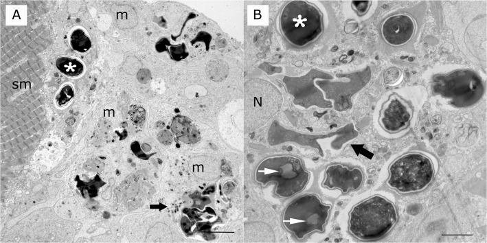
Fig 3. Transmission electron microscopy of microsporidian granulomatous myocytis in a yellow perch.
A) Multiple mature spores (asterisk) at various stages of digestion (wide arrow) and cell and parasitic debris inside macrophages (m) and a muscle cell (sm). Scale bar is 5 μm. B) Multiple mature spores (asterisk) at various stages of digestion are inside parasitophorous vacuoles and phagolysosomes of macrophages (wide arrow). Spores display a spore wall and posterior vacuoles (white arrow). Nucleus (n) of macrophage. Scale bar is 5 μm.

