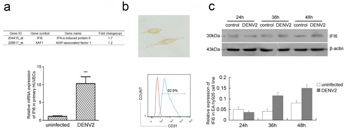
Fig 1. Dengue virus infection induced IFI6 over-expression in isolated primary HUVECs and EA.hy926 cells.

Expression of IFI6 was detected by GeneChip hybridization and analyzed(see Panel a, top) and mRNA expression of IFI6 was detected using qRT-PCR (see Panel a, below) at 48 hrs post-infection in HUVECs. Factor VIII antigen staining by immunocytochemistry (×400)(see Panel b, top). Analysis of CD31 expression in EA.hy926 cells by flow cytometry. The red was the negative control(see Panel b, below). (c) Protein expression of IFI6 was detected using immunoblotting in EA.hy926 cells after 24, 36, and 48 hrs post DENV2 infection. The gray scale scanning data were shown below and normalized to β-actin.
