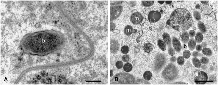
Fig 3. TEM micrographs of Ixodes ricinus pre-vitellogenic oocyte after feeding on guinea pigs infected with F. tularensis subsp. holarctica.
The morphology of some inclusions is typical of a Gram-negative bacterium, and their size is congruent with that of bacteria from the Francisella genus. m: mithocondrium, b: bacterium. Scale bar: A 0.22 μm, B 1.1 μm.

