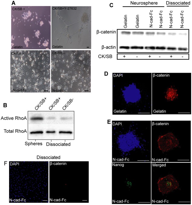
Fig 5. Suppression of Rho/ROCK and β-catenin signaling pathways by N-cadherin substrate in singly dissociated cells.
iPSC-derived neurosperes grown in suspension with and without CK/SB were enzymatically dissociated at day 5 and cultured on N-cadherin and gelatin substrate for 48 h. (A) In contrast to N-cadherin substrate, cells on gelatin failed to form neurites in the absence of Y-27632, as analyzed by bright-field images. Cellular phenotype on N-cadherin substrate was similar irrespective of the presence and absence CK/SB. (B) Compered to spheres seeded on N-cadherin substrate, pull-down assay shows down-regulation of active RhoA in dissociated cells. (C) β-catenin expression was analyzed by western-blotting. Immunostaining images for the expression of β-catenin (red) in shepres plated on (D) gelatin, and (E) N-cad-Fc substrate. β-catenin (red) expression is higher in Nanog expressing cells (green). (F) β-catenin expression was undetectable in dissociated cells plated on surfaces pre-coated with N-cad-Fc. DAPI shows total nuclei in the field of view. Scale bar: 50 μm.

