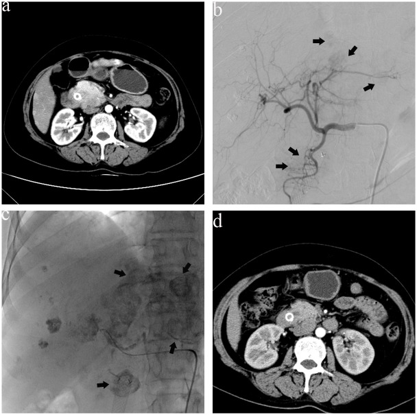
Fig 2. A 69-year-old man with a poorly differentiated adenocarcinoma of the middle third of the bile duct suffered stent blockage five months after stent implantation.
(a) One month after intraductal RFA, CT showed the tumor was enlarged with obvious enhancement, plus liver metastases. (b) Hepatic artery angiography revealed multiple metastases nodules in the liver (arrows). (c) After the second TACE, lipiodol depositions were found in the tumor (arrows) and liver metastases. (d) CT images obtained at 3-month follow-up showed the tumor had reduced in size.

