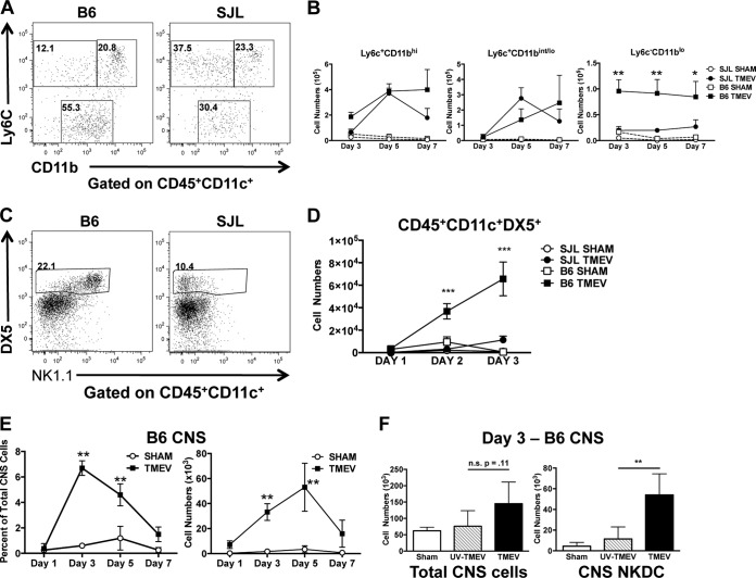
FIG 1 .
CNS viral infection results in accumulation of NK1.1-expressing dendritic cells in the CNS of TMEV-IDD-resistant B6 mice. Wild-type B6 and SJL/J mice were i.c. infected with 5 × 10⁶ PFU of TMEV or sham infected. Brains were harvested during acute infection, and mononuclear cells were isolated and stained for flow cytometry. (A) Live CD45+ CD11c+ cells were analyzed for expression of Ly6c and CD11b. Representative flow plots from day 5 post-TMEV infection are shown. (B) Numbers of cells in the CNS expressing Ly6C and CD11b on days 3, 5, and 7 p.i. in TMEV- or sham-infected B6 and SJL/J mice are shown. (C) Live CD3− CD11c+ cells were analyzed for their expression of NK1.1 and DX5. Representative flow plots from day 3 post-TMEV infection of B6 and SJL/J mice are shown. (D) Quantification of CD3− CD11c+ DX5+ cell numbers on days 1, 2, and 3 p.i. in TMEV- or sham-infected B6 and SJL/J mice. (E) Quantification of CD3− CD45+ CD11c+ NK1.1+ DX5+ brain cells on days 1, 3, 5, and 7 p.i. in TMEV- or sham-infected B6 mice are shown. (F) Quantification of total CNS lymphocytes and NKDCs in sham-infected (black bars), UV-inactivated TMEV-infected (hatched bars), or WT TMEV-infected (black bars) B6 mice 3 days p.i. Data are representative of 2 to 3 independent experiments with 3 to 5 mice per group. Error bars show standard deviations. ***, P < 0.001; **, P < 0.01; *, P < 0.05.

