FIG 2 .
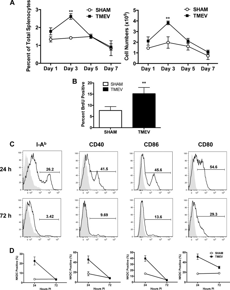
CNS TMEV infection induces proliferation and activation of splenic NKDCs. Wild-type B6 mice were i.c. infected with 5 × 10⁶ PFU of TMEV or sham infected. Spleens were harvested during acute infection, and mononuclear cells were isolated and analyzed by flow cytometry. (A) Quantification of CD3− CD45+ CD11c+ NK1.1+ DX5+ spleen cells on days 1, 3, 5, and 7 p.i. in TMEV- or sham-infected B6 mice. (B) At 24 and 48 h p.i., mice were i.p. injected with 1 mg BrdU in 100 µl sterile PBS. At 96 h p.i., the percentage of BrdU-positive NKDCs from spleens of TMEV- or sham-infected mice was quantified. (C) Spleens were harvested from B6 mice at 24 and 72 h post-TMEV infection, and expression levels of I-Ab, CD40, CD86, and CD80 on CD3− CD45+ CD11c+ NK1.1+ DX5+ spleen cells were determined. (D) Quantification of I-Ab-, CD40-, CD86-, and CD80-positive CD3− CD45+ CD11c+ NK1.1+ DX5+ spleen cells on days 24 and 72 h p.i. in TMEV- or sham-infected B6 mice. For histogram overlays, the shaded portion represents the isotype control, and the black line represents antibody staining. Data are representative of 2 to 3 independent experiments with 5 mice per group. Error bars show standard deviations. **, P < 0.01; *, P < 0.05.

