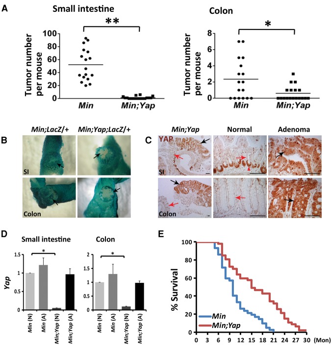
Figure 5.
YAP is required for intestinal tumorigenesis in APCMin/+ mice. (A) Quantification of the number of adenomas in the small intestines and the colons of 17 APCMin/+;Yapflox/flox (Min) and 18 VilCre;APCMin/+;Yapflox/flox (Min;Yap) mice at the age of 3 mo. (*) P < 0.005; (**) P < 0.001, t-test. (B) β-Galactosidase staining of Rosa26LacZ reporter in VilCre;APCMin/+;Rosa26LacZ/+ (Min;LacZ/+) and VilCre;APCMin/+;Yapflox/flox;Rosa26LacZ/+ (Min;Yap;LacZ/+) intestines. Note the blue (LacZ-positive) adenomas in the small intestines (SI) and colons of control Min;LacZ/+ mice and the white (LacZ-negative) adenomas in the colon and small intestine of Min;Yap;LacZ/+ mice. Black arrows indicate adenomas. (C) YAP staining in the rare adenomas in the small intestines (SI) and colons of Min;Yap mice. Note the absence of YAP staining in the nonneoplastic crypts (red arrows) and positive YAP staining in the neoplastic epithelia (black arrows). The red arrowhead indicates nonspecific staining of the antibody in the paneth cells in the ilea. Bar, 100 µm. (D) Real-time PCR analysis of Yap mRNA levels in normal tissues (N) and adenomas (A) from the small intestines and colons of APCMin/+; Yapflox/flox(Min) and VilCre;APCMin/+; Yapflox/flox (Min;Yap) mice. Note the sustained expression of Yap in Min;Yap adenomas but not Min;Yap normal tissues. Data are mean ± SD. n = 3. (*) P < 0.005, t-test. (E) Significantly prolonged life span in the Min;Yap mice compared with the control Min mice. Forty-two APCMin/+;Yapflox/flox (Min) and 47 VilCre;APCMin/+;Yapflox/flox (Min;Yap) mice were used for analysis. (Mon) Months.

