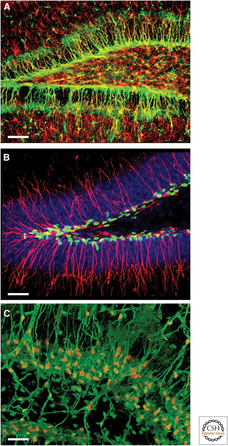
Figure 1.
Neural stem and progenitor cells are visualized in Nestin-fluorescent protein (FP) reporter mice. (A) Neural stem cell and progenitor cells (green) in the dentate gyrus (DG) of Nestin-green fluorescent protein (GFP) mice (glial fibrillary acidic protein [GFAP] staining for astrocytes, red). (B) Nuclei of neural stem cell and progenitor cells (green) in the DG of Nestin-CFPnuc mice (Nestin staining of radial processes, red; DAPI staining of the cell nuclei, blue). (C) Nuclei (orange) and soma of neural stem cell and progenitor cells (green) in the DG of the Nestin-GFP/Nestin-CFPnuc hybrid mice (images courtesy of JM Encinas, A-S Chiang, and G Enikolopov). Scale bars, 50 µm (A); 40 µm (B); 30 µm (C).

