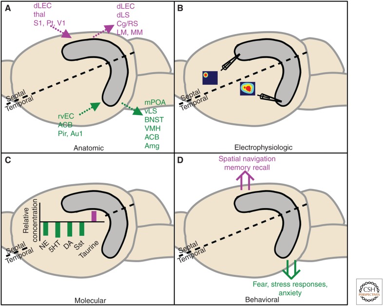
Figure 1.
Segregation of the hippocampus along the septotemporal axis. Septal (purple) and temporal (green) subregions of the hippocampus can be dissociated anatomically, electrophysiologically, molecularly, and behaviorally. (A) Dotted arrows represent anatomic efferents and afferents. ACB, Nucleus accumbens; Amg, amygdala; Au1, auditory cortex; BNST, bed nucleus of the stria terminalis; Cg/RS, cingulate/retrosplenial cortex; dLEC, dorsolateral entorhinal cortex; dLS, dorsolateral septum; LM, lateral mammillary nucleus; MM, medial mammillary nucleus; mPOA, medial preoptic area; Pir, piriform cortex; Pt, parietal cortex; rvEC, rostroventral entorhinal cortex; S1, somatosensory cortex; thal, thalamus; V1, visual cortex; vLS, ventro lateral septum; VMH, ventromedial hypothalamus. (B) Representative spatial place cell firing maps are shown in cartoon form, demonstrating a sharper receptive field in the septal and lower spatial resolution in the temporal hippocampus. (C) Relative levels of norepinephrine (NE), serotonin (5HT), γ-aminobutyric acid (GABA), glutamate (Glu), dopamine (DA), and somatostatin (Sst) are higher in the temporal hippocampus, whereas taurine is expressed at a higher level in the septal hippocampus. (D) Double arrows depict putative dissociations in behavioral functions along the longitudinal axis.

