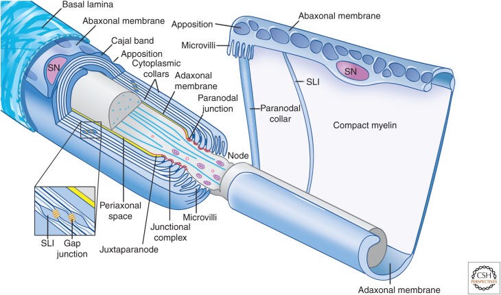
Figure 1.
Organization of myelinating Schwann cells. Schematic organization of myelinating Schwann cells (blue) surrounding an axon (gray); the left cell is shown in longitudinal cross section and the right cell is shown unwrapped. Myelinating Schwann cells are surrounded by a basal lamina (illustrated only on the left), which is in direct contact with the abaxonal membrane. The abaxonal compartment contains the Schwann cell nucleus (SN); it is divided into Cajal bands and periodic appositions that form between the abaxonal membrane and outer turn of compact myelin. The Schwann cell adaxonal membrane is separated from the axonal membrane by the periaxonal space (shown in yellow). Compact myelin is interrupted by Schmidt–Lanterman incisures (SLI), which retain cytoplasm and are enriched in the gap and other junctions; a similar autotypic junctional complex of adherens, tight and gap junctions, forms between the apposed membranes of the paranodal loops. Also shown are the paranodal loops and junctions (red) and the Schwann cell microvilli contacting the axon at the node. The axon diameter is reduced in the region of the node and paranodes. (This figure is adapted, with permission, from an original figure in Salzer 2003; modified in Nave 2010.)

