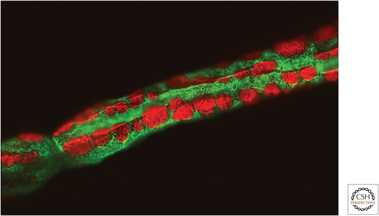
Figure 2.
Cajal bands. Fluorescence micrograph showing a portion of a teased, myelinated fiber from adult rat sciatic nerve, the presence of the Cajal bands (green), and the membrane appositions (red) in the abaxonal compartment. Staining shown is for phospho-NDRG1 (green) and α-dystroglycan (red) (see Heller et al. 2014 for further details).

