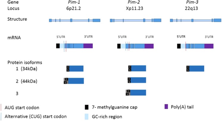
Figure 1.
Genetic structure of the Pims. The Pim kinases share significant homology (>60%).18 Each Pim gene contains 6 exons (depicted in darker blue). Pim mRNA contains a 5' untranslated region (UTR) which is comprised of a 7 methyl-guanine cap and GC-rich region which renders the Pims ‘weak transcripts' requiring cap-dependent translation.26 The 3' UTR contains destabilizing AUUUA motifs which result in a short Pim mRNA half life.25 Pim AUG start codons are located at nucleotides 431–433 and result in translation of one and two longer isoforms of Pim-1 and Pim-2, respectively.19 The longer 44kDa isoform of Pim-1 is derived from use of an upstream CUG start codon at nucleotides 158–160 and localizes to the plasma membrane, with a role in chemotherapeutic resistance.19 Pim proteins are autophosphorylated at an upstream serine 8 residue. A threonine residue and two downstream serine residues are also present. There is no regulatory domain and the overlapping catalytic and ATP-binding domains constitute the majority of the Pim proteins.

