. 2015 Jun 25;27(3):158–166. doi: 10.11919/j.issn.1002-0829.215036

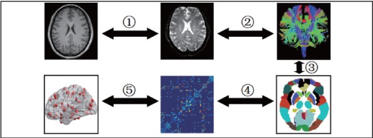
Figure 2. Flowchart of brain white matter (WM) structural network construction for patients with schizophrenia and healthy controls.

The weighted brain WM structural network for each participant is constructed using the following steps: (1) The T1-weighted image is registered a) to the corresponding baseline non-diffusion-weighted (b=0) image using a linear transformation and b) to the standard FA template in the Montreal Neurological Institute (MNI) 152 standard space (MNI-152 space) using the FSL non-linear transformation. Using the transformation matrices created in these two register processing steps, the resulting transformation matrix from diffusion space to MNI space is calculated and stored for later use. (2) The white matter fiber bundles in the whole brain are defined by using DTI deterministic tractography with a Diffusion toolkit (<ext-link ext-link-type="uri" xlink:href="http://www.trackvis.org">http://www.trackvis.org</ext-link>) to generate the three-dimensional curves characterizing cortical fiber tract connectivity and to reconstruct white matter tracts of the brain network based on the diffusion map. (3) An automated anatomical labeling (AAL) template is used to parcellate the brain into 90 cortical regions (45 for each hemisphere), in which each cortical or subcortical region represents a node of the cortical network. (4) The resulting weighted matrix is constructed by calculating the mean FA values of the connected streamlines between regions by the average of FA values of all voxels on each fiber track as the weights of the network edges. (5) The weighted brain WM structural network is constructed for each participant.