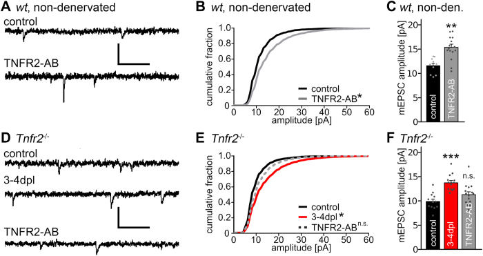
Figure 2. Denervation-induced homeostatic synaptic strengthening is observed in granule cells of TNFR1- or TNFR2-deficient slice cultures.
(A,D) Sample traces of whole-cell voltage-clamp recordings from slice cultures prepared from wild type and TNFR2-deficient mice. Cultures were denervated and recorded after 3–4 days post lesion (dpl) or were not denervated and treated for 3 days with TNFR2 activating antibodies (TNFR2-AB; scale bars: vertical, 20 pA; horizontal, 100 ms). (B,C) Non-denervated wild type slice cultures were treated with TNFR2-AB for 3 days [controls: n = 10 cells (pooled data from Fig. 1E); TNFR2-AB: n = 15 cells; from 4–6 cultures; in C: Kolmogorov-Smirnov test; in B: Kruskal-Wallis-test followed by Dunn’s post hoc test; *indicates p < 0.05; **indicates p < 0.01]. (E,F) TNFR2-deficient granule cells (Tnfr2−/−) showed an increase in mEPSC amplitudes after denervation. In non-denervated cultures the activation of TNFR2 with the TNFR2-AB did not lead to an increase in mEPSC amplitudes, thus confirming the specificity of the activating antibody (controls: n = 12 cells; 3–4 dpl: n = 13 cells; TNFR2-AB: n = 15 cells; from 5–6 cultures; in E: Kolmogorov-Smirnov test; in F: Kruskal-Wallis-test followed by Dunn’s post hoc test; *indicates p < 0.05; ***indicates p < 0.001; n.s., not significant).

