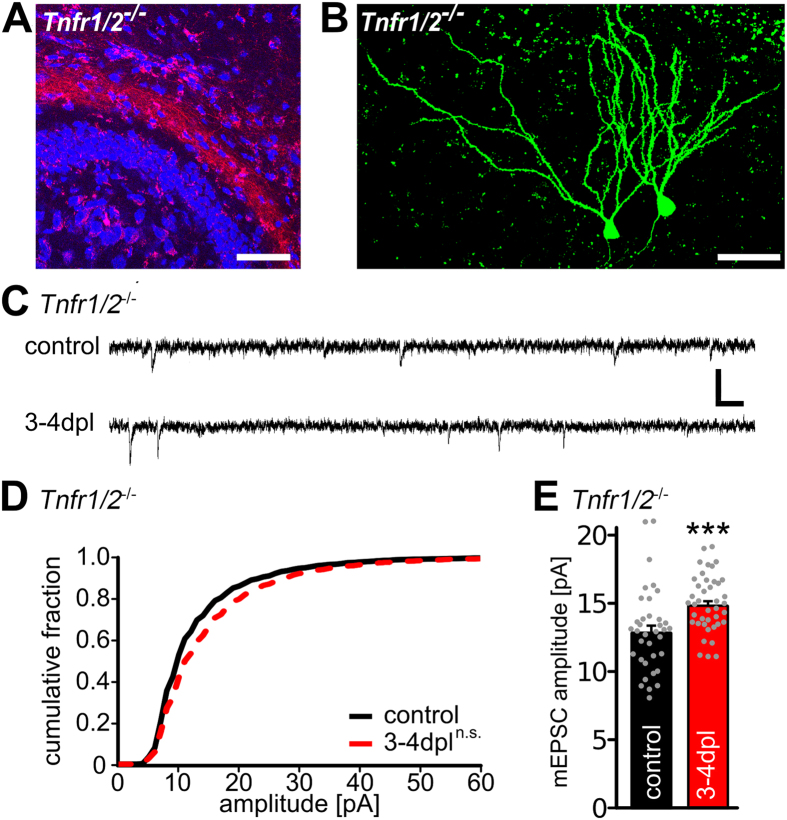
Figure 3. Granule cells deficient for both TNFRs show an impaired denervation-induced increase in synaptic strength.
(A) Mini-Rubi tracing of entorhino-hippocampal axons in TNFR1/2-deficient slice cultures (Tnfr1/2−/−). Entorhinal fibers terminate in the outer molecular layer of the dentate gyrus (blue: TO-PRO® nuclear staining, red: Mini-Rubi, scale bar: 100 μm). (B) Granule cells were patched with biocytin containing internal solution and stained with Alexa488-streptavidin (green) following the recording (scale bar: 50 μm). (C) Sample traces of whole-cell voltage-clamp recordings from granule cells of Tnfr1/2−/− slice cultures. (D,E) Cumulative distributions and mean values of mEPSC amplitudes. Recordings were made from control and denervated (3–4 dpl) slice cultures (controls: n = 36 cells; 3–4 dpl: n = 41 cells, from 15–17 cultures in D: Kolmogorov-Smirnov test; in E: Mann-Whitney-test; ***p < 0.001; n.s., not significant). See also supplementary information.

