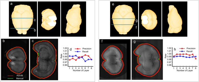
Figure 1. The effective reconstruction derived from RSVM.
(a) The reconstructed brain surface using RSVM shown in different views: horizontal plane, coronal plane and sagittal plane, respectively. (b,c) A comparison of the reconstructed boundaries of two cross-sections (labeled by the dark green lines in (a)) derived from RSVM (red) and manually (green). (d) 8 cross-sections from the dataset in (a) were selected for a quantitative evaluation; the recall and precision rate are calculated by comparing the reconstructions derived from RSVM with the manual reconstructions. (e–h) The analysis of the other brain dataset reveals the same information as (a–d).

