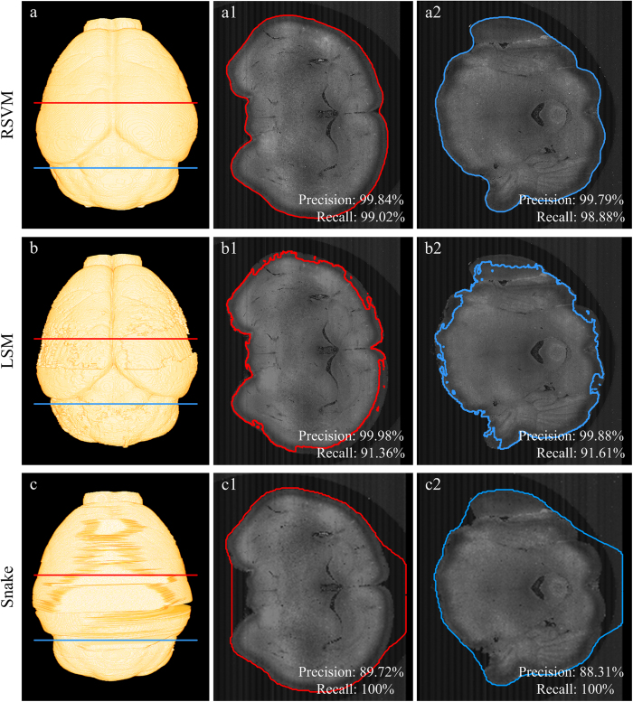
Figure 5. A comparison of the reconstructions derived from RSVM, the level set based method (LSM) and the active contour method (Snake).
(a–c) The mouse brain surface reconstructed by RSVM, LSM and snake, respectively. (a1)–(c1) and (a2)–(c2) The boundaries of a single coronal plane obtained through RSVM, LSM and snake, respectively. (a1)–(c1) correspond to the red line in (a–c), and (a2)–(c2) correspond to the blue line in (a–c). The recall and precision rates are given on the according extracted cross-sections in (a1)–(c1) and (a2)–(c2).

