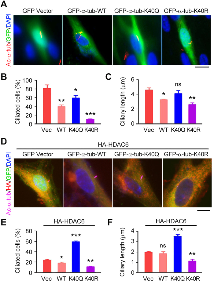
Figure 6. Deacetylation of α-tubulin is required for HDAC6-mediated ciliary disassembly.
(A–C) Immunofluorescence images (A), percentage of ciliated cells (B), and ciliary length (C) in RPE1 cells transfected with GFP vector or GFP-α-tubulin wild-type (WT), K40Q, or K40R, serum-starved for 24 hours, and stained with acetylated α-tubulin antibody and DAPI. Scale bar, 5 μm. (D–F) Immunofluorescence images (D), percentage of ciliated cells (E), and ciliary length (F) in RPE1 cells transfected with HA-HDAC6 and GFP vector or GFP-α-tubulin WT, K40Q, or K40R, serum-starved for 24 hours, and stained with HA and acetylated α-tubulin antibodies and DAPI. Scale bar, 5 μm. *P < 0.05, **P < 0.01, ***P < 0.001; ns, not significant. Error bars indicate SEM.

