Figure 1.
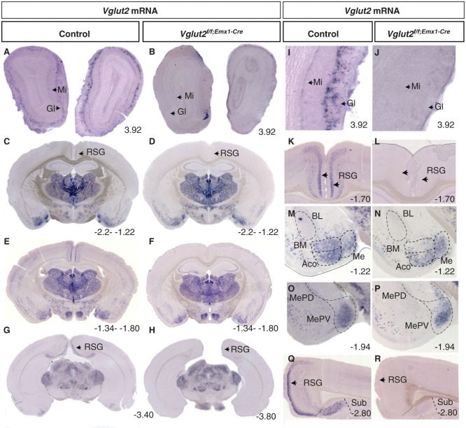
Specific deletion of Vglut2 in selected forebrain target areas. Floating in situ hybridization on coronal brain (70 μm) sections from control mice and Vglut2f/f;Emx1-Cre cKO mice (A–H) using a DIG-labelled Vglut2 probe. Close-ups as indicated in I–R, which demonstrate that cells expressing Vglut2 mRNA was absent in the mitral cell (Mi) layer and GI layer in the olfactory bulb (I–J). Vglut2 mRNA was also absent in the RSG of the medial cortex in the Vglut2f/f;Emx1-Cre mice (K, L). There is a loss of Vglut2 mRNA in the BMA, in the ACo the mRNA is partially deleted, and the Me amygdala and MePV are unaltered in the Vglut2f/f;Emx1-Cre mice (M–P). Vglut2 mRNA positive cells were present in the Sub in control mice but not in Vglut2f/f;Emx1-Cre cKO mice (Q, R). ACo = anterior cortical amygdaloid area; BL = basolateral amygdaloid nucleus; BM = basomedial amygdaloid nucleus anterior part; cKO = conditional knock-out; DIG = digoxigenin; GI = periglomerular layer; Me = medial amygdaloid nucleus; MePV = posteriorventral medial amygdaloid nucleus; Mi = mitral cell layer; RSG = retrosplenial group; Sub = subiculum; Bregma interval (dorsal, ventral) is shown in lower right corner.

