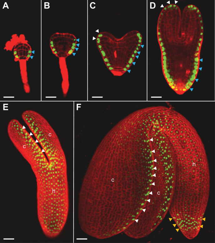
Fig. 3.
Dynamic expression of CLE19 during embryogenesis. As examined under a confocal microscope in embryos excised from pCLE19:SV40-3XGFP:tCLE19 transgenic plants, GFP expression was first observed in protodermal cells (indicated by blue arrowheads) at the lower portion of the 32-cell stage embryo (A), and persisted in their progeny cells in the triangular-stage embryo (B). Additional GFP expression was observed in epidermal cells at the tips and abaxial sides of the cotyledon (indicated by white arrowheads) in heart-shaped (C) and torpedo-stage embryos (D). In walking-stick (E) and cotyledonary-stage embryos (F), strong GFP expression was seen in epidermal cells located at the edges of cotyledons (c, indicated by white arrowheads) and in root caps (indicated by yellow arrowheads), weak GFP expression was observed in the hypocotyl (h). (E) and (F) were prepared by superimposing multiple scanned images. Scale bars=50 μm.

