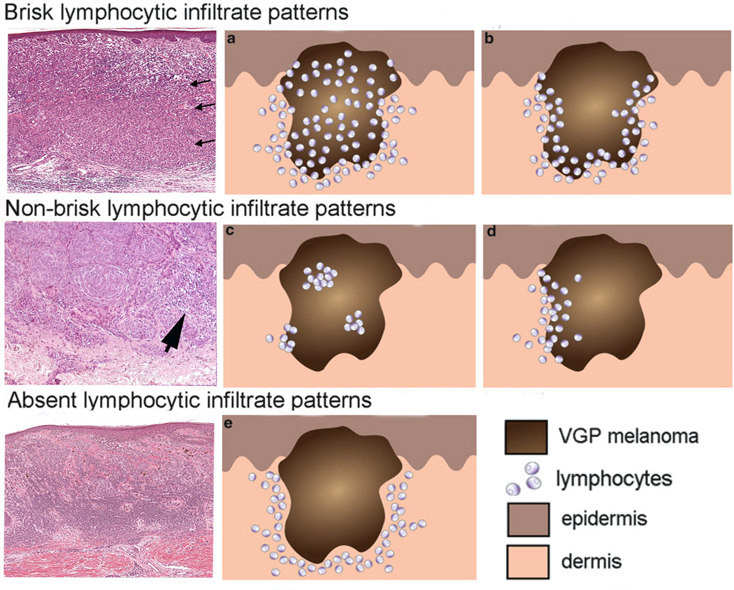
Figure 3.
Brisk, non-brisk, and absent lymphocytic infiltration in primary melanoma nodules. The three photomicrographs in the left column exhibit the brisk, non-brisk and absent patterns. The brisk pattern is defined by lymphocytes present throughout the tumor as shown by arrows in the top left panel. Non-brisk is defined by scattered, focal groups of lymphocytes (arrow). Panel 3: Absent is defined by no lymphocyte infiltration in the tumor. Panels (a, b, c, d, e) in the middle and right columns are schematic renderings of different types of TIL patterns in primary melanoma. The brisk lymphocytic response may involve the entire tumor nodule (panel a) or be present along the advancing edge of the entire peripheral nodule (panel b). The focal nature of the non-brisk pattern is demonstrated in panel c and d. In the rendering of the absent lymphocytic response, there are either no lymphocytes in the tumor, or if lymphocytes are present (panel e) they do not interact with melanoma cells. Panels a, b, c, d, e are adapted from figure 4 of chapter 16 by Schatton and colleagues, entitled “Tumor Infiltrating Lymphocytes and their Significance in Melanoma Prognosis,” in Molecular Diagnostics for Melanoma. Thurin M, Marincola FM eds; Humana Press. 2014, 287–324.

