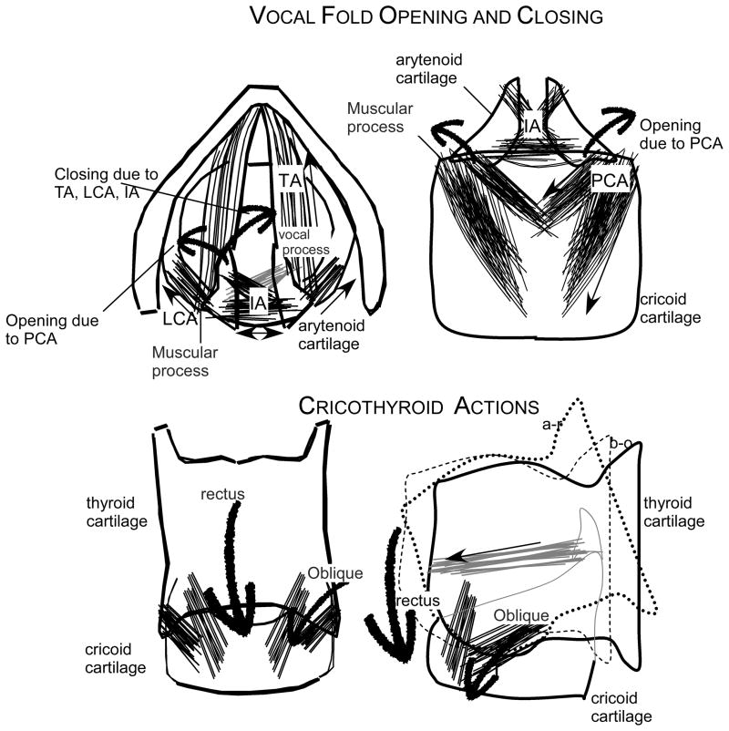
Figure 1.
Schematic illustration of the laryngeal muscles, LCA is the lateral cricoarytenoid, TA is the thyroarytenoid, IA is the inter-arytenoid and the PCA is the posterior cricoarytenoid, as well as the rectus and the oblique compartments of the cricothyroid muscle. The location of the arytenoid cartilage with the muscular and vocal processes, the cricoid cartilage and the thyroid cartilage. In the top left diagram the motion of the vocal process forward and downwards during closing of the vocal folds due to the actions of the adductor TA, LCA and IA muscles and the trajectory of the vocal process upwards and lateral with contraction of the PCA. In the bottom 2 illustrations, the effects of contraction of the rectus and oblique compartments of the cricothyroid muscle pull the thyroid cartilage forward (oblique) and downwards (rectus) to lengthen the vocal fold. This is adapted from Figure 1 of Ludlow (Ludlow, 2005).

