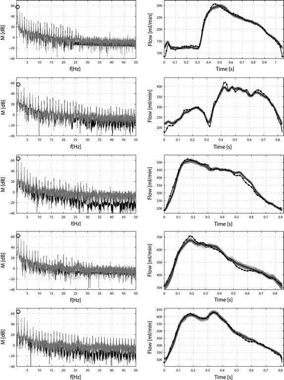
Fig. 9.

Comparison between flow rates calculated by the Womersley’s solution in in vivo blood vessels and in the in vitro test bench. The Fourier spectral analysis was calculated in order to assess the differences in reproduced frequencies between Womersley’s solution (black line) and ones obtained in vitro (grey line) (left-hand panel). On that graph the circle point out the fundamental frequency of the heart cycle. The right panel is presented flow rate obtained using the in vitro test bench. The solid black line with points presents desired signal, the solid grey line presents measured cycle, solid black line presents average of 60 measured cycles. Rows from top to bottom show patients from 1 to 5.
