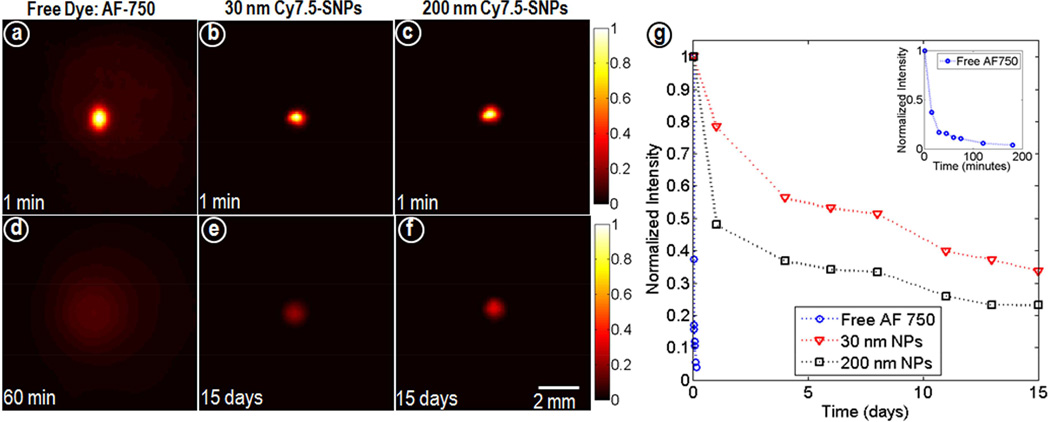
Figure 2.
Fluorescence images of agarose phantoms (n=3) acquired at different time points for (a, d) AF-750*, (b, e) 30nm and (c, f) 200 nm Cy7.5-SNPs and (g) normalized intensity profiles. All acquisition parameters were kept constant and normalized to the maximum at minute 1. *Hydrophilic dye AF-750 was used as free dye as Cy7.5 gets quenched in aqueous media.

