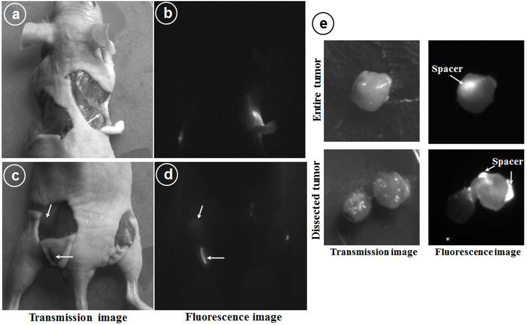
Figure 5.
Optical fluorescence images of the dissected mice 14 days post spacer implantation. (a&b) upper flank to show left Cy7.5-SNPs-spacer and right Cy7.5-spacer, (c&d) hind flank: on left Cy7.5-SNPs-spacer implanted intradermally and right side was implanted intratumorly; (e) Dissected tumor from the mice to locate the implanted spacer inside tumor (upper panel) and lateral section to visualize nanoparticles distribution (lower panel).

