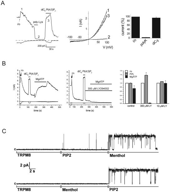
Figure 2.
Activation of TRPM8 excised inside-out patches and planar lipid bilayers. A. Representative trace for recording of TRPM8 currents in large patches in Xenopus oocytes with 500 μM menthol in the patch pipette from (Rohacs et al., 2005). Left panel shows currents at −100 and +100 mV, from ramp protocol shown in the middle. Application of 30 μg/ml poly-Lysine and 50 μM diC8 PI(4,5)P2 are indicated with the horizontal lines. B. Similar excised inside out patch measurement from (Yudin et al., 2011), demonstrating the effect of 2 mM MgATP and LY294002. Right panel shows summary for 300 μM LY294002 that inhibits both PI4K and PI3K, and for 10 μM LY294002 that selectively inhibits PI3K. C. Representative measurement in planar lipid bilayers with purified TRPM8 from (Zakharian et al., 2009). In the upper trace first the TRPM8 protein shows no activity in the absence of menthol and PI(4,5)P2, then diC8 PI(4,5)P2 is applied, then menthol, in the continuous presence of PI(4,5)P2. In the bottom trace menthol and PI(4,5)P2 is applied in the reverse order.

