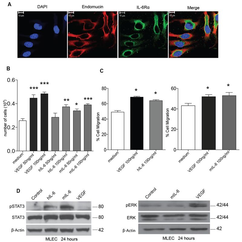
Figure 2. IL-6 has direct effects on mouse lung endothelial cells.
A. Immunocytochemistry staining for endomucin (red) and IL-6Rα (green) on MLEC. B. 70,000 MLEC were plated and treated with either PBS (control) and indicated concentrations of VEGF or IL-6 for 72 hours. The cells were then trypsinized and counted using a cell counter and the mean of the triplicates were calculated. Proliferation assay shows a significant increase in proliferation with VEGF (50 or 100ng/ml), human IL-6 (100ng/ml) and mouse IL-6 (50 or 100ng/ml). C. Scratch assay using a time-lapse microscope was used to measure the migration of MLEC after treatment with VEGF and IL-6. Significant increases in cell migration are observed with VEGF or IL-6 treated MLEC after 16 hours. Statistical analysis carried out using student T-test is shown as (*) p ≤ 0.05: (**) p ≤ 0.01; (***) p ≤ 0.001 D. Western blot analysis of protein extracted from MLEC treated with IL-6 (30ng/ml) or VEGF (30ng/ml) for 24 hours. IL-6 induced downstream pSTAT3 and VEGF induced downstream pERK levels, indicating both pathways are independent of each other for signaling in MLEC.

