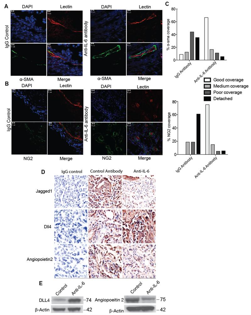
Figure 5. Effects of anti-IL-6 antibody on in vivo IGROV-1 vasculature.
1×10⁷ IGROV-1-luc cells were injected i.p. into 8 weeks old Balb/c nude female mice. After 24 hours the mice were treated with either control IgG antibody (20mg/kg) or anti-IL-6 antibody (20mg/kg) twice a week for 4 weeks. Following this, three of the animals from each group were injected with TRITC-conjugated Bandeiraea via the tail vein, 3 min before being perfused with 4% paraformaldehyde. The lectin stained frozen sections were stained for α-SMA (green) (A) or NG2 (green) (B). Treatment with anti-IL-6 antibody was able to restore the pericyte coverage in IGROV-1 xenografts. Staining of α-SMA and NG2 staining show poor or detached pericyte coverage in the IgG antibody group compared to good pericyte coverage in the anti-IL-6 treated group. C. Quantification of the staining show a significant difference between the IGROV-1 antibody control and anti-IL-6 treated group (p value < 0.001). The quantification carried out by two independent reviewers was analyzed using chi-sqaure test (n=6 per group from two independent experiments). D. Immunohistochemistry staining for Jagged1, DLL4 and Ang2 in the IGROV-1 xenograft following 4 weeks of treatment with anti-IL-6 antibody. The images were taken from 5 randomly selected areas per tumor section (n=6 per group from two independent experiments) using a ×40 microscope. E. Western blot analysis of DLL4 and Ang2 from protein extracted from in vivo IGROV-1 xenografts treated with anti-IL-6 antibody.

