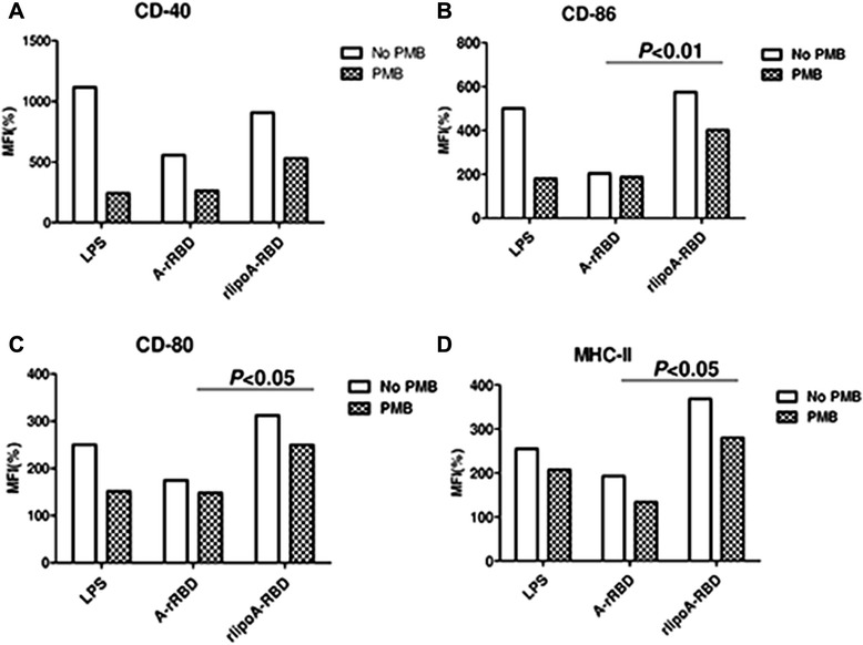
Fig. 3.

Up-regulation of surface biomarkers of BMDC by rlipoA-RBD. BMDC from C57BL/6 was collected and treated with GM-CSF on days 0 and 3. A-rRBD and rlipoA-RBD were treated on day 6 for 18 h, then DC were collected to analyze their surface markers, including CD-40 (a), CD-86 (b), CD-80 (c), and MHC-II (d) by flow cytometry. All groups were divided into polymyxin B (PMB) treated (black-net bar) or without (white bar) to validate insignificant LPS contamination. All surface marker signaling was normalized by calculating the ratio of mean fluorescence intensity (MFI) between medium control and treatments. The experiments had been performed at least three times
