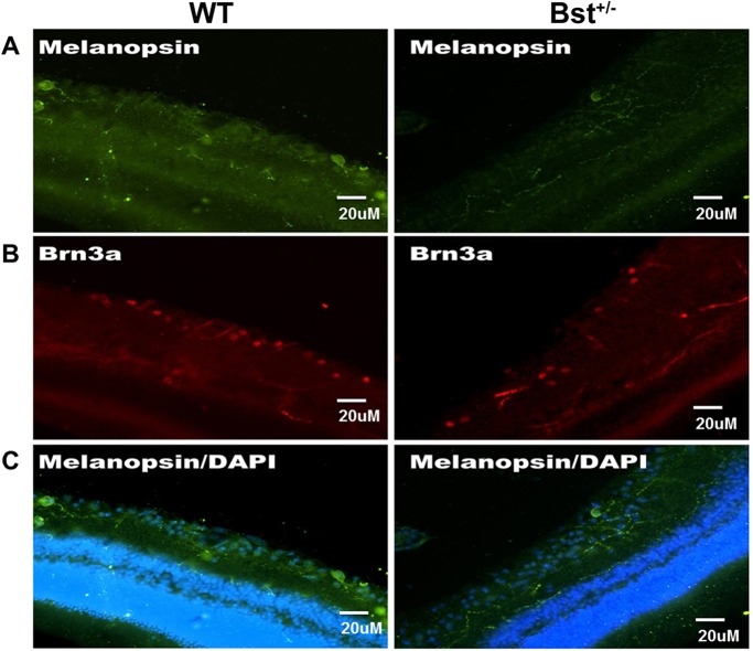
Fig. 3.
Characterization of RGCs in WT and Bst+/− retinas. Sections of WT and Bst+/− mice retinas were stained with anti-melanopsin and anti-Brn3a antibodies, and images were taken by using the same confocal microscopy parameters for primary antibody. (A) Melanopsin+ RGCs (arrow) in retinas harvested from Bst+/− and WT mice. (B) There were fewer Brn3a+ RGCs in Bst+/− retinas than in WT retinas. (C) Superimposed melanopsin (green) immunolabeling with DAPI nuclear counterstain (blue).

