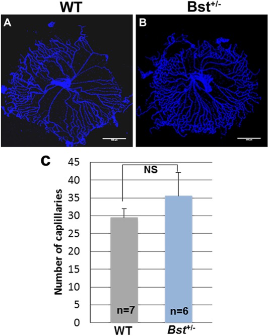
Fig. 5.

Delayed hyaloid regression in Bst+/− mice. Hyaloid vessel in retinas collected from WT (A) and Bst+/− mice (B) at postnatal day 8. (C) Quantitative analysis of capillaries. A circle was drawn around the hyaloid prep and the number of vessels crossing a concentric circle with half the radius was counted. Error bars are s.e.m. NS, not significant.
