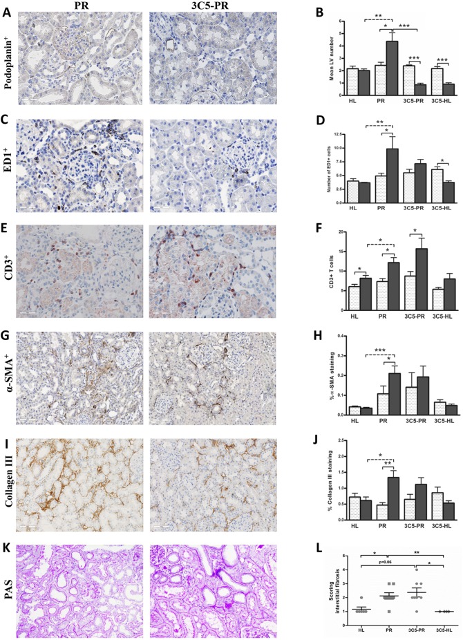
Fig. 2.
Effects of anti-VEGFR3 antibody treatment on renal lymphangiogenesis, inflammation and fibrosis. Quantification of renal cortical podoplanin-positive vessel-like structures of the rats who received treatment with anti-VEGFR3 antibody (IMC-3C5) showed a significant reduction of LV number at 12 weeks (A,B; 400×), whereas it showed a non-significant trend in reducing the number of macrophages in the cortical interstitium of proteinuric rats (C,D; 400×), and did not influence T-cell influx (E,F; 400×) at week 12. Anti-VEGFR3 antibody also did not have a significant effect on α-SMA (G,H; 200×), collagen III deposition (I,J; 200×) and interstitial fibrosis (K,L; 200×). White dotted bars represent week 6 before treatment; black bars represent week 12 after treatment. The PAS staining quantification (interstitial fibrosis) is showed at 12 weeks. HL, healthy; PR, proteinuric untreated; 3C5, anti-VEGFR3 antibody. *P<0.05, **P<0.01, ***P<0.001.

