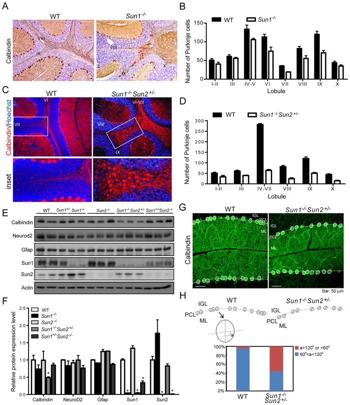
Fig. 4.
Aberrant positioning of Purkinje cells in Sun1-null cerebellum. (A) Cerebellum from 30-day-old mouse stained for calbindin (brown) by immunohistochemistry. Some calbindin-positive cells were found in the white matter of Sun1−/− cerebellum (indicated by red arrows). Numerals indicate the lobules. (B) Quantification of Purkinje cells in each sagittal section at the surface of the IGL in WT and Sun1−/− cerebellum. Statistics (mean±s.e.m.) were obtained from independent measurements of four mice for each genotype. (C) Calbindin (red) in 30-day-old mouse cerebellum was stained using immunofluorescence. Many calbindin-positive cells were found in the white matter of Sun1−/−Sun2+/− cerebellum (boxed, inset). DNA was stained by Hoechst 33342 (blue). (D) Quantification of Purkinje cells in each sagittal section at the surface of the IGL in WT and Sun1−/−Sun2+/− cerebellum. Statistics (mean±s.e.m.) were obtained from independent measurements of four mice for each genotype. (E) Western blot analysis for protein expression of calbindin, Neurod2, Gfap, Sun1 and Sun2 in cerebellums of adult mice with the indicated genotypes. (F) Quantification of the relative protein expression levels shown in E. Statistics are mean±s.e.m. *P<0.05, Student's t-test. (G) Purkinje cells of 30-day-old WT and Sun1−/−Sun2+/− cerebellums were immunostained for calbindin (green). The Purkinje cell bodies are outlined with white ovals. The white dashed lines mark the border between the Purkinje cell layer (PCL) and the inner granule layer (IGL). ML, molecular layer. (H, upper scheme) Schematic presentation of the alignment of the Purkinje cell soma shown in G. The angle (indicated as ‘a’) of the long axis of the soma, and the border (dashed line) between the IGL and ML was measured for each Purkinje cell. (Lower graph) Quantification of ‘a’ in 50 Purkinje cells of each genotype. 96% of WT and 44% of Sun1−/−Sun2+/− Purkinje cells showed ‘a’ between 60° and 120°; and 4% of WT and 56% of Sun1−/−Sun2+/− Purkinje cells showed ‘a’ >120° or <60°. Scale bars: 50 μm.

