Fig. 4.

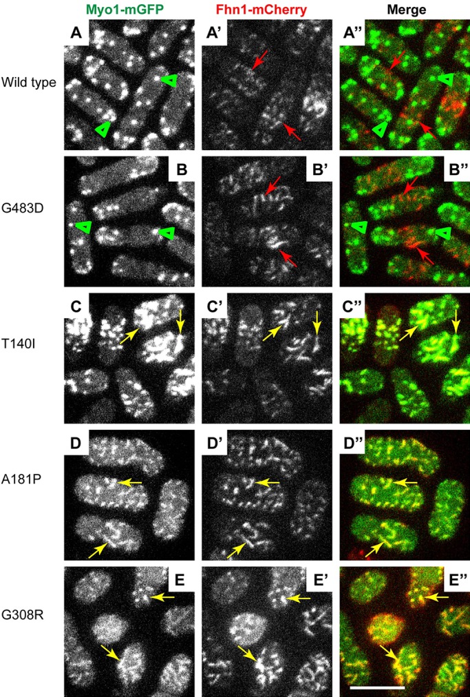
Myo1 motor domain mutants colocalize with the eisosome marker Fhn1. Confocal images show the localization of (left panels, A-E) mGFP-tagged (A) wild-type or (B-E) mutant Myo1, (middle panels, A′-E′) mCherry-tagged Fhn1 (Fhn1-mCherry), and (right panels, A″-E″) merge of Myo1-mGFP and Fhn1-mCherry images. The wild-type and Myo1(G483D) localize to actin patches (green arrowheads) that are distinct from Fhn1-mCherry in eisosomes (red arrows). The T140I, A181P and G308R Myo1 mutants colocalize with Fhn1 in eisosomes (yellow arrows). The images represent maximum intensity projections of three consecutive optical sections through the top surface of the cell acquired at 0.4-µm intervals. Scale bar: 10 µm.
