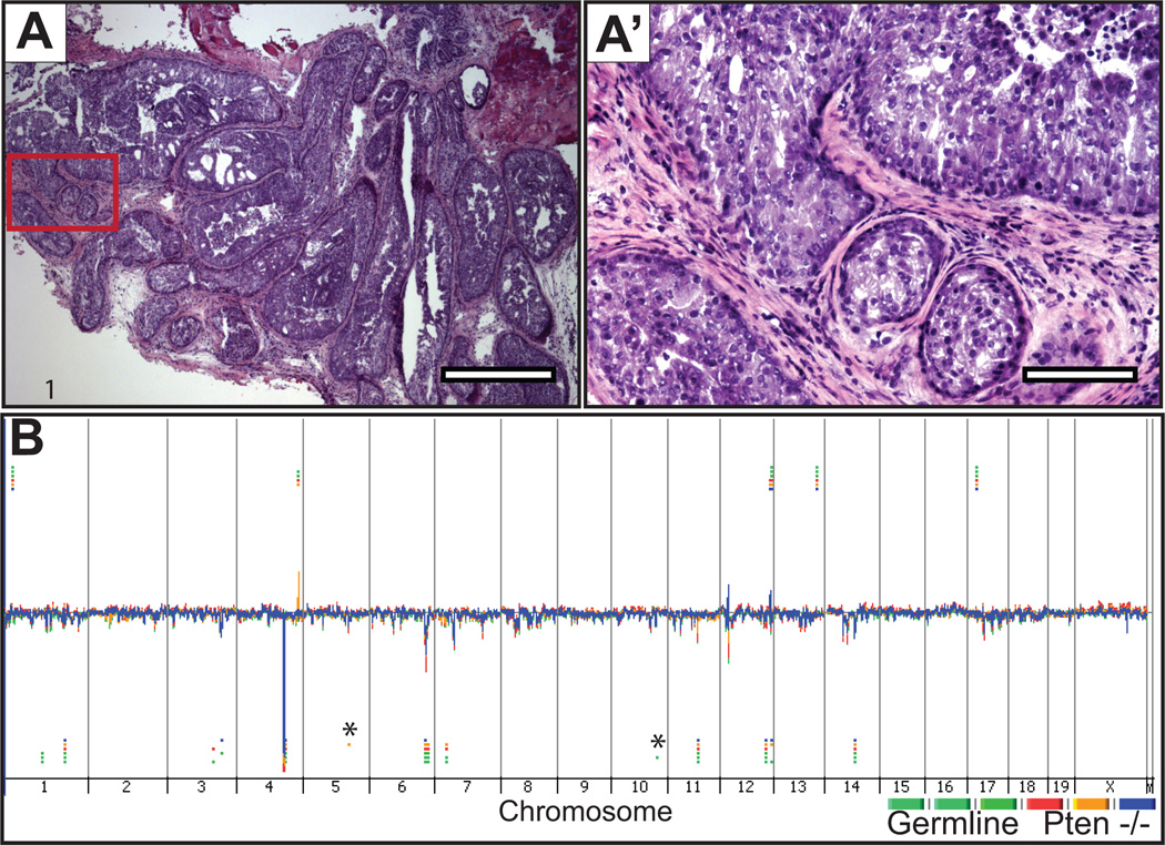
Figure 2. Histology and genomic view of prostates from Pten null (Pb-Cre+;PtenL/L) mice.
A-A’, representative microscopic images from hematoxylin and eosin-stained sections of prostate glands from which DNA was extracted for CGH hybridization shown at low (4×) and high-power (20×) magnification, respectively. B, Genomic DNA from Pb-Cre+;PtenL/L prostate tumors and germline spleen were hybridized against sex-matched normal mouse C57BL/6 reference DNA using Agilent CGH slides containing 180K probes. Whole genome view of overlaid moving averages (2 Mb window) for the log2 ratios of fluorescence between a sample/reference DNA probe (Y axis) plotted at its genomic position (X axis), red, blue and yellow (tumors; n = 3) and green shades (germline n = 3). Aberrations called by the ADM-2 algorithm are identified by horizontal bars. (*) Aberrations called by the ADM-2 algorithm in a single sample, that by manual inspection exhibited similar fluorescence intensities in all samples including germline and tumor (see Supplemental Figure S4, for representative examples). (1) CNAs in common between germline, and Pten lesions that are not visually obvious in the genome view window.

