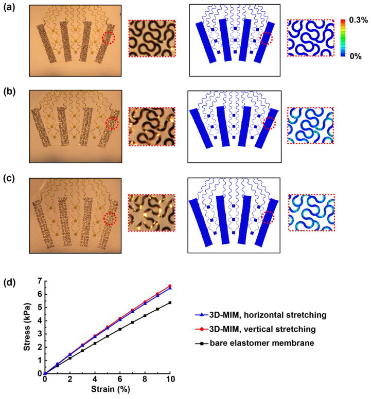
Figure 2.
(a–c) Optical images (left) and FEA results of the distribution of maximum principal strain (right) of a representative device (a) in the undeformed state, (b) with 20% uniaxial stretching in the horizontal direction, and (c) with 15% biaxial stretching. The insets show magnified views of the fractal electrode. (d) FEA results of the stress-strain relation of the elastomer membrane and the 3D-MIM.

