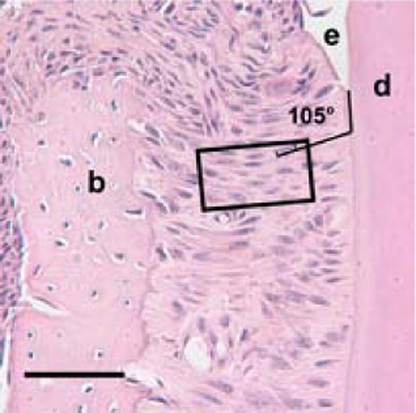
Fig. 1.

Measurement area within the cervical periodontal ligament of a coronally sectioned day 41 (dpn) CD44 wild-type mouse molar, × 40. The rectangle outlines the sampling region for angular measurements of periodontal ligament fibroblasts. Lines define orientation of the cervical root region relative to a fibroblast cell nucleus within the sampling region, as 105°. Enamel is labeled (e), dentin (d) and alveolar bone (b). Scale bar = 50μm.
