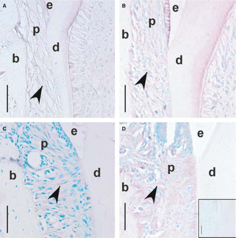
Fig. 2.

CD44 immunostaining of periodontal ligament fibroblasts in the cervical region during mandibular first molar (M1) eruption and occlusion in CD44 wild-type mice. Coronal sections of M1 at days 11 (A), 14 (B), 18 (C) and 26 (D; dpn), × 40. Reddish stain (arrowheads) shows staining for CD44 within the periodontal ligament. Methyl green counterstain shows labeling of cell nuclei. Inset box (D) shows the negative control, day 26 (dpn), and an absence of CD44 immunostaining. Enamel is labeled (e), dentin (d), bone (b) and the periodontal ligament (p). Scale bar = 50μm.
