
Fig. 5.
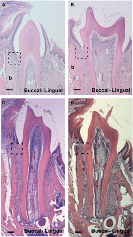
Coronal sections of mandibular first molars (M1) showing CD44 wild-type and knockout mice during preocclusal eruption, day 18 (dpn; A, B) and early occlusion, day 26 (dpn; C, D), × 10. Dotted-line square highlights the developing periodontal ligament shown in Fig. 4. Scale bar = 100μm.